Recent techniques such as confocal optical microscopy and high-resolution scanning electron microscopy have opened up a new perspective on these structures by providing us with three-dimensional views of their architecture.
|
Recent techniques such as confocal optical microscopy and high-resolution scanning electron microscopy have opened up a new perspective on these structures by providing us with three-dimensional views of their architecture. |
|
Cyanobacteria and prochlorophyta are technically bacteria, and have a bacterial cell organization, but possess all the photosynthetic chemistry of green plants.
Some cyanobacteria live in caves, where they survive on very little light and may play a significant part in precipitating calcite on stalactites and stalagmites. With Julia James, Bill Allaway, Neville Michie and Anya Salih I have been studying these using confocal microscopy, electron microscopy and physiological methods.
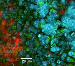
On the Great Barrier Reef, and on other coral reefs around the world, cyanobacteria and their relatives, the prochlorophyta, live in symbiosis with sponges and ascidians (sea-squirts). The prochlorophytes are particularly interesting from the point of view of cell evolution since they seem to be long-lost descendants of the ancestor of the chloroplast of green plants. With Tony Larkum and Teresa Dibbayawan I have been looking at the ultrastructure of both prochlorophytes and symbiotic cyanobacteria on coral reefs.
 |
|
 |
|
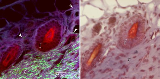
The novel technique of second harmonic imaging provides a high resolution and extremely sensitive method of studying the three-dimensional distribution of collagen in tissue.